


A perspective by M.J. Aman in mBio profiles our work to image how Ebola virus fuses and escapes into host cells in real time. Read our paper here.
VIDEO CAROUSEL


FEATURED NEWS
Featured


LATEST NEWS
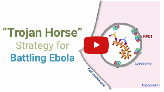
Fatal Attachment: Watch the Ebola virus fusion machine in action inside a human cell
/Like a Trojan horse, Ebola virus sneaks into a human cell by hiding out inside lysosomes. In order to escape from the lysosome, the virus must then fuse its membrane with the membrane of the lysosome. Live imaging captures this moment of fusion in brilliant color. Images by Chandran Lab postdoctoral fellow, Dr. Jennifer Spence.
This Week in Evolution (TWiEvo) podcast→
/
In episode 5 of the weekly podcast, "This Week in Evolution," Kartik Chandran and Sara Sawyer join hosts Nels Elde and Vincent Racaniello to talk about how the filovirus receptor NPC1 regulates ebolavirus susceptibility in bats.
Read the paper here and listen below.
Catching Ebola virus in the Act of Fusion→
/
The American Society of Microbiology (ASM) profiles our work to image how Ebola virus fuses and escapes into host cells in real time. Read our paper here.
Study Reveals Arms Race Between Ebola Virus and Bats, Waged for Millions of Years→
/
Our study in eLife reveals an evolutionary arms race between Ebola virus and bats, waged over many millions of years. Read the press release and our blog post.
Novel Hantavirus Infection Method→
/The Scientist reports on our paper in mBio describing the highly cholesterol-dependent hantavirus cell entry mechanism.
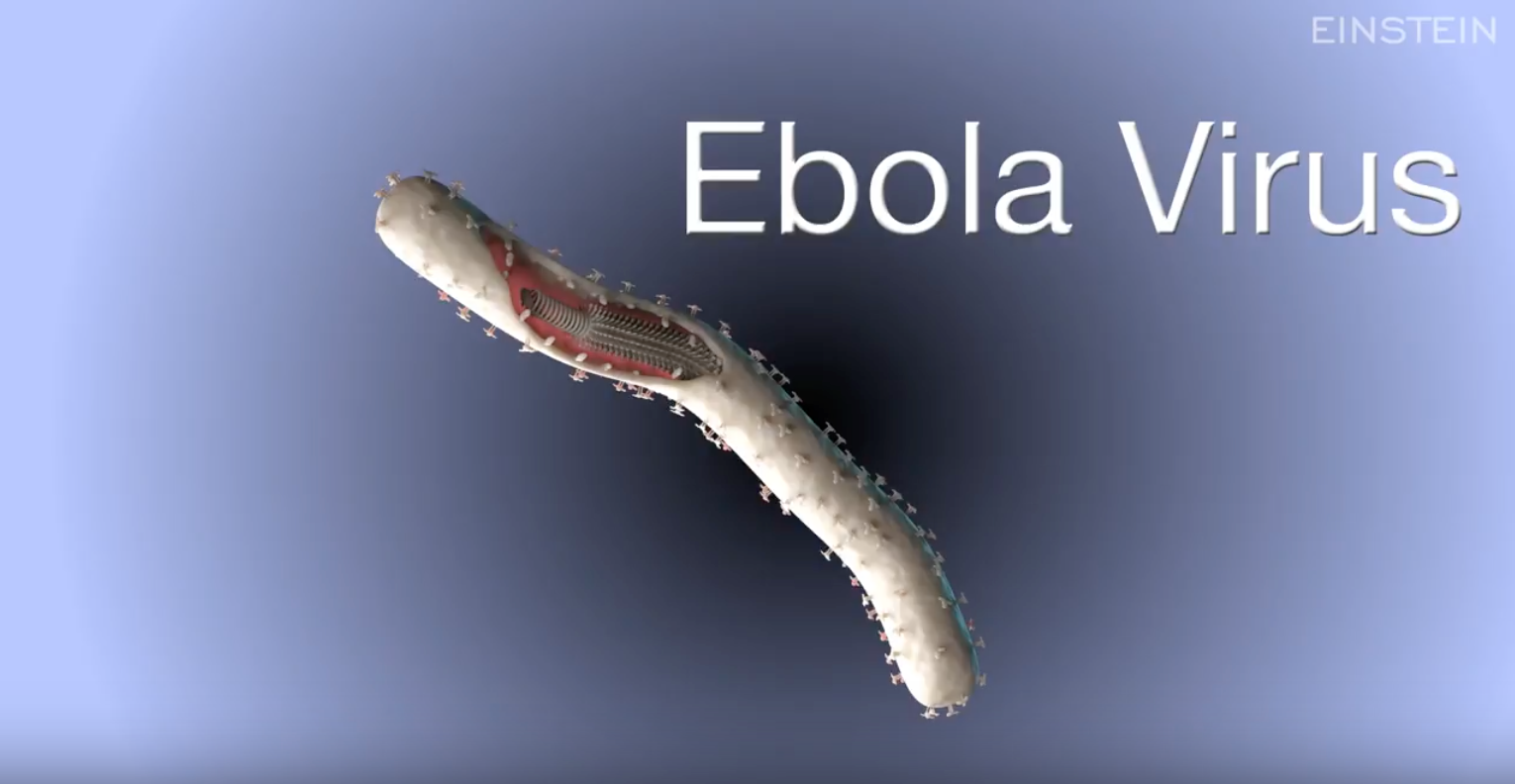
How Ebola infects a cell
/This 3D animation shows how Ebola virus exploits a naturally occurring protein in our cells called NPC1 to cause infection and spread in the body. Narrated by Kartik.
Study identifies Ebola virus's Achilles' heel→
/
ScienceDaily.com discusses our mBio paper, which showed that NPC1 is indispensable for Ebola virus infection, replication, and disease in a mouse model.
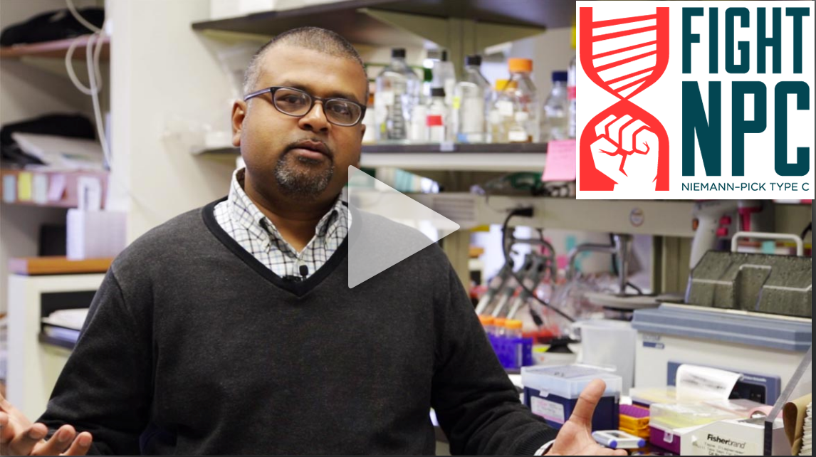
The Ebola-NPC1 Link→
/
Fight NPC, an initiative dedicated to advocacy and support for Niemann-Pick type C patients and families and NPC research, features our work on the Ebola-NPC1 link. Fight NPC supports research in the Chandran Lab that aims to better understanding this connection.
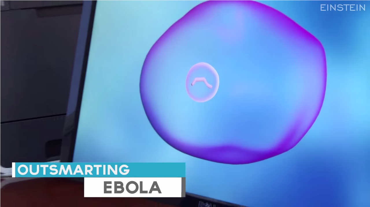
Outsmarting Ebola Virus: Innovative Research on Treatment
/Kartik explains the novel strategies he and his colleagues are using to develop treatments for Ebola virus infections. Watch 3D animations showing how Ebola enters a cell and delivers its payload in order to replicate and spread throughout the body.
Understanding the Ebola virus→
/
The Ebola epidemic in west Africa has highlighted the threat of emerging infectious diseases to public health. In this Nature.com webcast (registration required), Kartik outlines the importance of basic molecular research to understand how Ebola enters human cells, and why such research has been key to developing drugs and vaccines. Dr. Simon Hay (University of Oxford) also explains how mapping and modeling can help identify where are the places where Ebola and other emerging diseases risk jumping from animals to people. Watch the video (Kartik's talk only, 21 minutes).
Einstein Goes Viral
/Episode 314 of Dr. Vincent Racaniello's world-famous virology blog, This Week in Virology (TWiV), features Kartik and two other Einstein virologists, Margaret Kielian and Ganjam Kalpana. Filmed before a live audience in Einstein's Price Center Auditorium. Watch the video (1 hour, 26 mins).
Unraveling Ebola→
/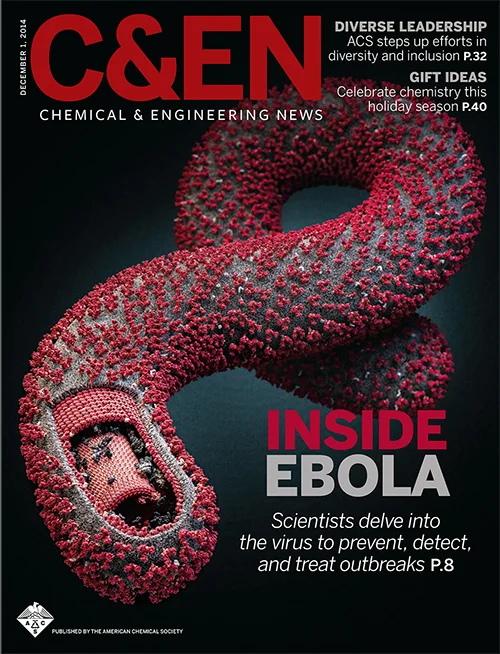
In this C&EN Magazine (Chemical and Engineering News) cover story, Kartik and other Ebola researchers and clinicians discuss efforts to decipher the inner workings of Ebola virus, current emergency drug candidates, and the development of next-generation anti-Ebola drugs.
The road to Ebola→
/
What prompted Kartik to study the entry mechanism of Ebola virus? Read this Science Careers article to find out more about why and how Kartik, Esther Ndungo, a graduate student in his lab, and several other researchers chose to investigate Ebola virus.
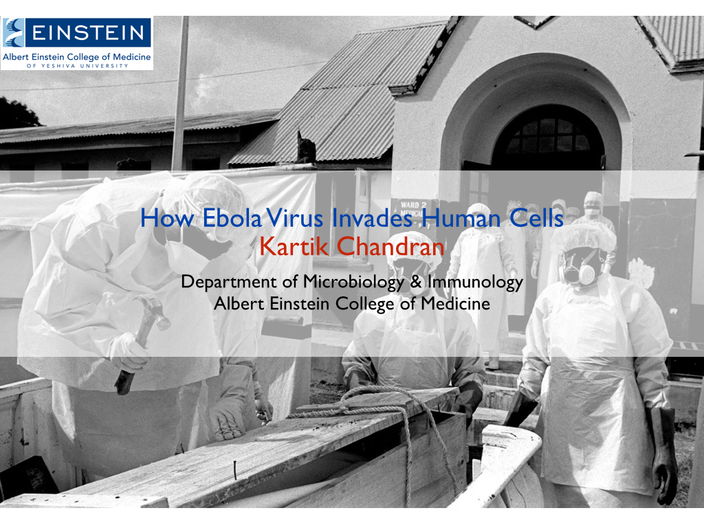
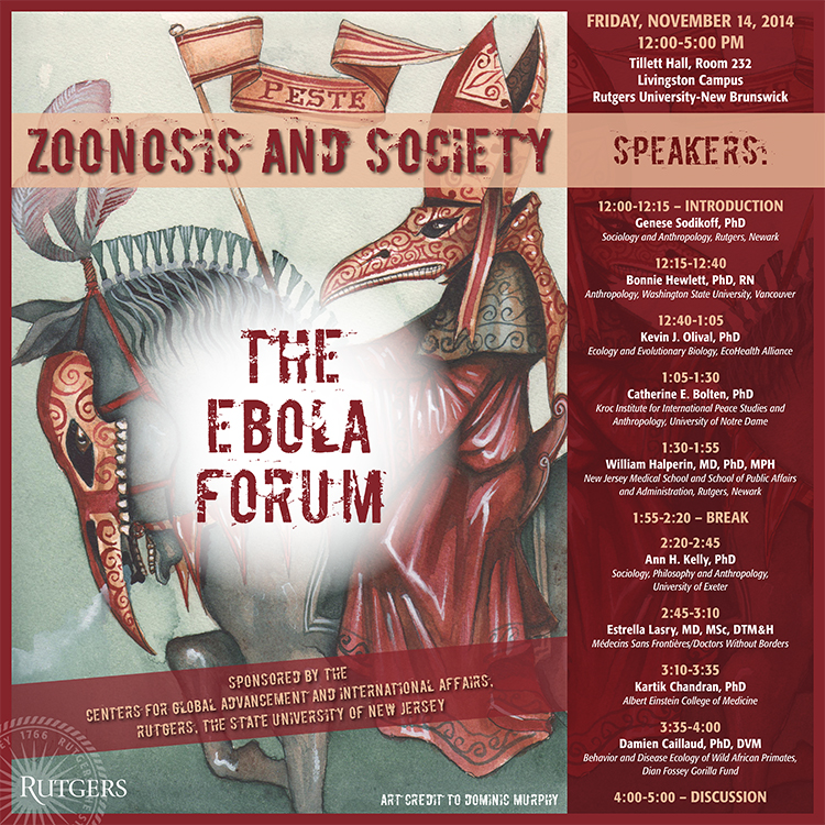
Seminar at The Ebola Forum, Rutgers University→
/Kartik spoke at this symposium organized by the working group, "Zoonosis and Society: Interdisciplinary Perspectives of Animal-to-Human Disease" at Rutgers University. Its goal was to integrate the diverse perspectives of sociologists, anthropologists, epidemiologists, ecologists, clinical researchers, and molecular virologists on the Ebola outbreak in West Africa. Watch the video of Kartik's talk (34 minutes).
Researchers Study Ebola Link to Gene in Rare Disease→
/The Wall Street Journal interviews Kartik and Chandran Lab collaborator Steven Walkley about the connection between the rare genetic disease Niemann-Pick Type C and Ebola virus. Our research raises the possibility that a mutated NPC1 gene offers protection against Ebola infection, just as carriers of sickle-cell disease are protected against malaria. (subscription only)
More coverage on this story at The Scientist and CBSNews.com.
Don’t fear flying during the Ebola crisis: experts→
/
The New York Daily News interviews Kartik about the evolving Ebola epidemic and the risk of airline travel in light of news that someone who later tested positive for the virus traveled by air while infected.
Experimental Drug Would Help Fight Ebola if Supply Increases, Study Finds→
/
The New York Times interviews Kartik about new research that found the experimental Ebola drug ZMapp was successful in treating monkeys infected with the virus.
Ebola Drug’s Success Bolsters Approach for Other Diseases→
/Bloomberg News speaks with Kartik about ZMapp, the experimental antibody treatment for Ebola, and passive antibody therapeutics for other viral infections.
The Race to Find a Vaccine or Treatment to Stop Ebola→
/
CBSNews.com interviews Kartik about the Ebola outbreak and his research on the virus. Kartik explains how Ebola enters cells, and how understanding this process has been instrumental in identifying new targets for drug development.